
dinoflagellate morphology
Dinoflagellates are morphologically distinct from other eukaryotes in the structure of their dinokont flagellation and their dinokaryotic nucleus = dinokaryon (Fensome et al. 1993; Hoppenrath
2017; Hoppenrath & Saldarriaga 2008 Tree of Life webpage for dinoflagellates).
dinokont flagellation = two dissimilar flagella: one ribbon-like transverse flagellum, with paraxial rod and simple hairs on the surface, in multiple waves beating to the cells’ left, and one more conventional, cylindrical to
flattened, longitudinal flagellum beating posteriorly
dinokaryon = permanently condensed chromosomes without typical eukaryotic histones, thus without nucleosomes, and with an extranuclear spindle that passes through cytoplasmic channels — closed dinomitosis
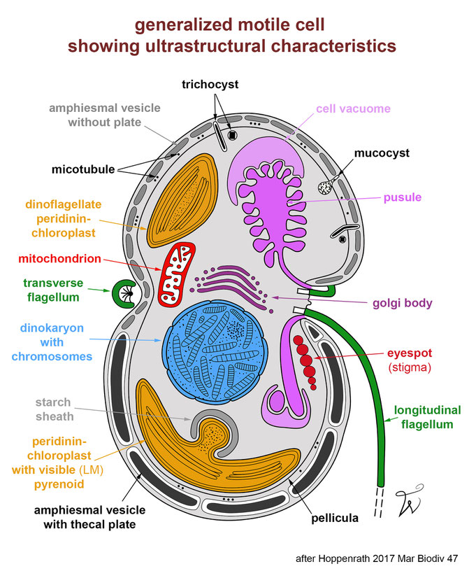
Hoppenrath 2017 (Mar. Biodiv. 47):
The amphiesma (= cortex or wall or periplast) consits of alveolar vesicles (= amphiesmal vesicles in dinoflagellates, which can contain cellulosic thecal plates) lying beneath the cell membrane (= plasmalemma or plasma membrane). Usually microtubules are present under the vesicles. In some taxa a continuous fibrous layer, the dino-pellicle, can be found under the alveolae. The flagella normally lie in grooves, the transverse one in the cingulum (= girdle) and the longitudinal one partly in the sulcus. In most dinoflagellates, the cingulum divides the cell into an upper and a lower part. The sulcus defines the ventral cell side. In some taxa no depressed cingulum exists (podolampids), and in prorocentroids the flagellation is desmokont (i.e., the typical dino-flagellation is not associated with grooves). A special organelle of dinoflagellates is the pusule, a special vacuole of still unknown function, open at the flagellar bases. Mitochondria have tubular cristae. Dinoflagellates can contain different types of extrusomes (i.e., organelles that secrete material to the exterior): trichocysts, mucocysts, taeniocysts, and/or nematocysts.
Do you want to get a more realistic 3D impression of a thecate photosynthetic dinoflagellate cell of Ensiculifera tyrrhenica? Please have a look at this movie showing a segmentation of a FIB-SEM dataset.
From Mocaer et al. 2023 J Cell Sci 136: doi:10.1242/jcs.261355
photo
©
K. Mocaer
explanatory pages are here:
- general cell terminology, cell orientation & flagellation page
- cingulum path page
- tabulation types page
- Kofoid system of tabulation nomenclature (plate naming) page
- apical structures page
- types of ornamentation page
- types of eyespot page
- extrusomes
- chloroplasts
SENCKENBERG am Meer
Deutsches Zentrum für Marine Biodiversitätsforschung
PD. Dr. Mona Hoppenrath
Südstrand 44, 26382 Wilhelmshaven
