
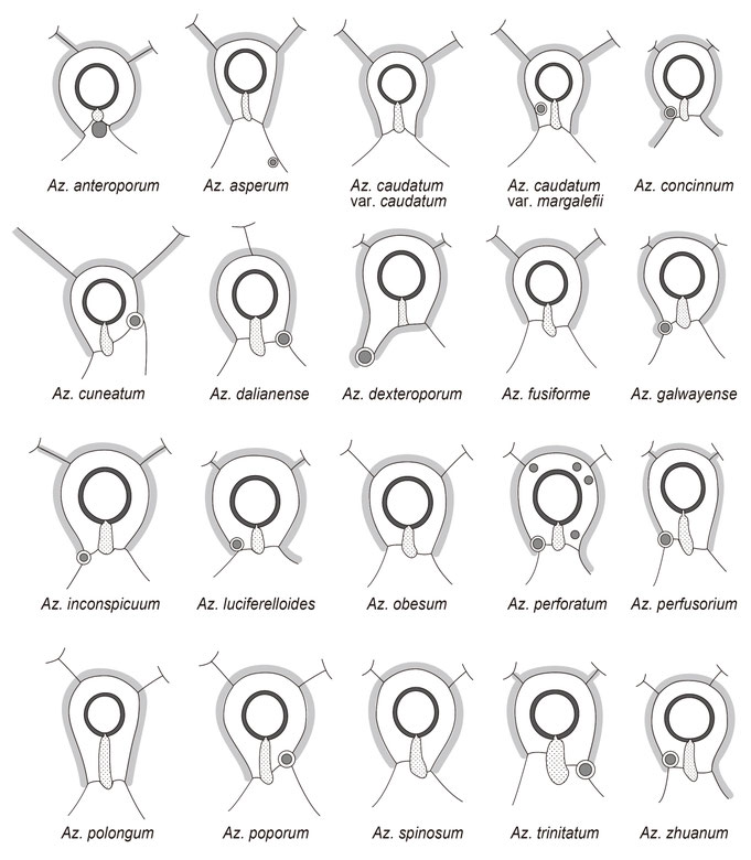
comparative drawings of Azadinium species
These drawings (to scale) illustrate the diversity of the main characters used to identify Azadinium species in the light microscope. Recognize the different cell sizes and shapes, the location of the nucleus and the number and location of pyrenoids.

The epithecal tabulation is species specific. Especially compare the number, shapes and sizes of the apical and anterior intercalary plates and the arrangement of precingular plates.

Also the microarchitecture of the apical pore complex (APC), especially the shape of the outer pore plate (Po) and the location of a ventral pore (vp) is species-specific.
SENCKENBERG am Meer
Deutsches Zentrum für Marine Biodiversitätsforschung
PD. Dr. Mona Hoppenrath
Südstrand 44, 26382 Wilhelmshaven
